It looks like you're using an Ad Blocker.
Please white-list or disable AboveTopSecret.com in your ad-blocking tool.
Thank you.
Some features of ATS will be disabled while you continue to use an ad-blocker.

The Human Brain research project
page: 2share:
biggie,
I have a TON of info on the Paranormal aspects and the use of "f"MRI, PET and CAT scans, If you need anything on "healing" give a hollar!!!
I am hoping for another interview with Dr. Menchen and would be happy to pass on any questions you may have to him.
He has some small experience in "Functional MRI's"
I am also hoping to obtain a few more interviews from some very prestigious individuals in the field to compliment Dr. Green, as well as assist in the research.
Let me know, I am a U2U away..
Semper
ps. Welcome to the Team.
S
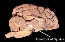
The Aqueduct of Sylvius
source for image: google.com/images
and thats the Aqueduct of Sylvius...
Aqueduct of Sylvius: A canal that communicates between the third and fourth ventricles in a system of four communicating cavities within the brain that are continuous with the central canal of the spinal cord.
The four ventricles consist of the two lateral ventricles, the third ventricle and the fourth ventricle:
* Lateral ventricles: The lateral ventricles are in the cerebral hemispheres. Each lateral ventricle consists of a triangular central body and four horns. The lateral ventricles communicate with the third ventricle through what is called the interventricular foramen (opening).
* The third ventricle is a median (midline) cavity in the brain that is bounded by the thalamus and hypothalamus on either side. Anteriorly (in front) the third ventricle communicates with the lateral ventricles and posteriorly (in back) the third ventricle communicates with the aqueduct of Sylvius (also called the aqueduct of the midbrain).
* The fourth ventricle is the most inferior (lowest) of the four ventricles of the brain. It extends from the aqueduct of the midbrain to the central canal of the upper end of the spinal cord with which it communicates by the two foramina (openings) of Luschka and the foramen (opening) of Magendie.
The ventricles are filled with cerebrospinal fluid, which is formed by structures called choroid plexuses located in the walls and roofs of the ventricles.
source: medterms.com
source for image: google.com/images
and thats the Aqueduct of Sylvius...
Prior to my posting information on "F"MRI's (Functional), here is some information on the MRI and how it works...
Next, "Functional" MRI and studies involving various activity in each area of the brain.
Semper
How does the procedure work?
MRI is a unique imaging method because, unlike the usual radiographs (x-rays), radioisotope studies and even CT scanning, it does not rely on radiation. Instead, radio waves are directed at protons, the nuclei of hydrogen atoms, in a strong magnetic field. The protons are first "excited" and then "relaxed," emitting radio signals, which can be computer-processed to form an image. In the body, protons are most abundant in the hydrogen atoms of water—the "H" of H2O—so that an MRI image shows differences in the water content and distribution in various body tissues. Even different types of tissue within the same organ, such as the gray and white matter of the brain, can easily be distinguished. Typically an MRI exam consists of two to six imaging sequences, each lasting two to 15 minutes. Each sequence has its own degree of contrast and shows a cross section of the head in one of several planes (right to left, front to back, upper to lower).
~~~~~~~
How is the procedure performed?
Magnetic Resonance Imaging (MRI) procedureThe patient is placed on a sliding table and a radio antenna device called a surface coil is positioned around the upper part of the head. After positioning the patient with the head inside the MRI gantry, the radiologist and technologist leave the room and the individual MRI sequences are performed. The patient is able to communicate with the radiologist or technologist at any time using an intercom. Also, many MRI centers allow a friend or, if a child is being examined, a parent into the room. Depending on how many images are needed, the exam will generally take 15 to 45 minutes, although a very detailed study may take longer. The patient will be asked not to move during the actual imaging process, but between sequences some movement is allowed. Patients are generally required to remain still for only a few seconds at a time. Some patients will require an injection of a contrast material to enhance the visibility of certain tissues or blood vessels. A small needle connected to an intravenous line is placed in an arm or hand vein. A saline solution will drip through the intravenous line to prevent clotting until the contrast material is injected about two-thirds of the way through the exam.
When the exam is over the patient is asked to wait until the images are examined to determine if more images are needed.
~~~~~~~
What are the limitations of MRI of the Head?
Bone is better imaged by conventional x-rays, and CT is preferred for patients with severe bleeding, acute trauma or who because of their medical condition are unable to tolerate an MR scan procedure. MRI may not always distinguish between tumor tissue and edema fluid and does not detect calcium when this is present within a tumor. In most cases the exam is safe for patients with metal implants but there are a few exceptions, so patients should inform the technician of an implant prior to the test. The exam must be used cautiously in early pregnancy. MRI often costs more than CT scanning.
www.radiologyinfo.org...
Next, "Functional" MRI and studies involving various activity in each area of the brain.
Semper
Hey guys, these are some links that should help you get acquainted with the basis of healing via intention (biokinesis if you will).
www.abovetopsecret.com...
www.abovetopsecret.com...
www.abovetopsecret.com...
So far I have been attuned in several forms of Reiki. I started with Kundalini, then Gold, Tacyon, and then Ethereal Crystals...There are other forms called Usui Reiki and I'm sure many others that I am ignorant of.
www.abovetopsecret.com...
www.abovetopsecret.com...
www.abovetopsecret.com...
So far I have been attuned in several forms of Reiki. I started with Kundalini, then Gold, Tacyon, and then Ethereal Crystals...There are other forms called Usui Reiki and I'm sure many others that I am ignorant of.
Biggie,
Why not post a comprehensive soliloquy on your experiences in this area and the possible benefits another may expect.
Semper
Why not post a comprehensive soliloquy on your experiences in this area and the possible benefits another may expect.
Semper
Broca's Speech Area, Wernicke's Area, Arcuate Faciculus
Broca's Speech Area is named after Pierre Paul Broca, who was the first to site this area of the brain in 1861.
Source: Wikipedia.org
the arcuate fasciculus connects Brocas area with Wernicke's area like so:
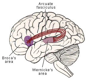
Wernicke's Area is located in the left temporal lobe, posterior to the primary auditory complex. It is responsible for language comprehension. See above pics. It was named after Karl Wernicke in 1874.
Broca's Speech Area is named after Pierre Paul Broca, who was the first to site this area of the brain in 1861.
Source: Wikipedia.org
Broca's area is the section of the human brain (in the opercular and triangular sections of the inferior frontal gyrus of the frontal lobe of the cortex) that is involved in language processing, speech production and comprehension. Broca's and Wernicke's areas are found unilaterally in the brain.
It comprises of Brodmann's Area 44,[1] and some authorities also include Brodmann's Area 45[2][3][4]); Broca's Area is connected to Wernicke's area by a neural pathway called the arcuate fasciculus. The corresponding area in macaque monkeys is responsible for high-level control over orofacial actions.[5]
source: wikipedia.org
the arcuate fasciculus connects Brocas area with Wernicke's area like so:
Wernicke's Area is located in the left temporal lobe, posterior to the primary auditory complex. It is responsible for language comprehension. See above pics. It was named after Karl Wernicke in 1874.
The benefits I have been introduced to thanks to the Reiki treatment are as follows:
Increased psychic ability
Increased connection to source
Heightened states of awareness that can be facilitated through breathing exercises or hand techniques
Clarity of thought for short periods of time (I have severe ADD, for me this is a god-send)
several others that I cannot think of at this current moment
The first attunement I received was quite shocking actually. I had a spiritual experience during this occassion where my whole body was filled with "fire" (possibly the holy spirit/higher self) that lifted me up off the ground (or so I felt) and then slowly dragged me back down to Earth. The only way I can describe this is as some kind of initiation into the mysteries that every great teacher has talked about (Egyptian mystery religions, Gnosticism, etc). I was initiated into the White Lodge a few months ago and since then my life has never been the same. If you wish to find out for yourself if these are truly authentic experiences, I suggest you have an attunement done yourself. The website you can request them is www.xehupatl.com. The man's name is Steven Kammerhofer and he is my teacher.
I hope everyone has a chance to discover natural healing that everyone has the power to do. However, you must first believe you are able to do so or none of the healing will work. Good luck my friends and may the true light guide you on your journey!
What could be fascinating, is to have you induced into that kind of elemental state during a "Functional" MRI...
This would provide the information necessary to determine the area of the brain most active during this experience.
Semper
This would provide the information necessary to determine the area of the brain most active during this experience.
Semper
I spoke with Dr. Mencken again and he eluded more completely his experience with Functional MRI's.
Apparently he was interning and assisting with a research project in which a subject was place is an "f"MRI and shown various images. The flow of blood was then observed traveling to various parts of the brain and thereby certain hypotheses were extrapolated as to brain function in relation to neural activity.
At one point during a specific day, a member of the research team developed a debilitating migraine. (note: for those that have true migraines, they are devastating.)
The research member was immediately rushed under the "f" MRI and a "fast" image done. This produced the result of indicating the area of the brain that was being effected by the migraine.
Now research has shown that migraines are different for different people, but this one action alone started researchers in a direction that is currently revealing fascinating new evidence in pain relief and effective treatments for migraines.
I will be following this up with actual MRI images of the subject.
Semper
AIR or "Automatic Image Registration" is ameliorating the "F"MRI process by potentially expediting the review process.
The obvious conclusion to the data is that as computer speed increases and processing time reduces, the research will take leaps forward and yield more productive results.
Semper
Functional MRI and AIR
Cohen and his colleagues are helping to shape a newly evolving discipline called cognitive neuroscience. In recent work, they have exploited technology developed during the 1980s that many scientists believe will revolutionize study of the brain. Imaging technology, such as magnetic resonance imaging (MRI) and other techniques, combined with computing power makes it possible, in effect, to peel away the bone and membrane surrounding the brain. Without even touching their human subject, researchers can see what happens inside a living, thinking brain, and they can identify what parts of this intricate, complexly folded, interconnected mass of tissue "light up" during mental activities.
Cohen and colleagues use a technique known as functional MRI to record a view of the functioning brain that is among the most detailed yet reported. While other brain-mapping techniques give what resembles a satellite view of the world, in which cities can be seen and identified, the Pitt/CMU researchers can see streets. With functional MRI, they can map the sites of brain activity to a resolution as fine as one millimeter, comparable to mapping a football field in six-inch units.
Cohen and his colleagues used a conventional MRI machine, like those that became available in many hospitals during the 80s, a big advantage since the research can be accomplished without major new investment in technology. Functional MRI works on the principle that when brain cells (neurons) become active, blood flows to them, and the MRI scanner registers increased oxygen in the area. Because MRI machines used in this way detect changes resulting from biological function, the method got its name.
The technique generates large amounts of data quickly -- a great advantage, says Cohen, and a problem. "It gives a lot of information to work with, but likewise it's a tremendous amount of data to process -- as much as half a gigabyte per experiment." To deal with the data overload, Cohen and his colleagues turned to the Pittsburgh Supercomputing Center's Alpha Cluster, a linked network of 14 DEC Alpha workstations. They used the cluster to address a particular problem of their functional MRI experiments. A human subject stays in the machine for two to three hours as the MRI scanner records data. Though special pillows are used to reduce movement, it's impossible to keep the head perfectly still. Software called automatic image registration (AIR), developed by Roger Wood of UCLA, can correct for head movement, but the sheer number of images -- typically 1,200 per experiment -- creates an imposing demand on computing.
AIR is an ideal application for the Alpha Cluster, notes Cohen, because it is inherently parallel. A single experiment typically records 200 separate images for each of 6 separate scan sites, or slices. "For each slice," says Cohen, "you take as a reference point one of the 200 images and align all the others to it. There's no need for communication back and forth. Each sample can be aligned to the reference independently."
On a high-speed workstation, says Cohen, it took as much as 24 hours computing time to register the images from one experiment. "Often, we run two experiments in an evening, which means the computing can't keep up with the data, and this cripples the research." On the Alpha Cluster, the same computing takes an hour, a radical speedup that overcomes the research bottleneck.
www.psc.edu...
The obvious conclusion to the data is that as computer speed increases and processing time reduces, the research will take leaps forward and yield more productive results.
Semper
"F" MRI computer imaging..
Interesting especially when compared with Tone's posting of the specific areas of the brain and their functions.
Semper
[edit on 10/29/2006 by semperfortis]
This image shows what regions in a subject's brain were involved in a memory task. This kind of study leads to improved understanding of "working memory", which affects clinical treatment of schizophrenia and amnesias.
www.psc.edu...
This 3-D image of a subject's brain shows that the primary visual cortex becomes activated while the subject, who is inside an MRI scanner, looks at the whirling pattern.
www.psc.edu...


Interesting especially when compared with Tone's posting of the specific areas of the brain and their functions.
Semper
[edit on 10/29/2006 by semperfortis]
A graphic and simplistic representation of the process..

Semper
Semper
The Physiological activities involved with "Working Memory."
Closer and closer we are coming to understand each section of the brain and it's functions in regards to our physiology.
Semper
A light changes from green to yellow, and you jam your foot on the brake. It's almost an automatic impulse, a no-brainer, you might say, guided by the knowledge, planted somewhere, that moving-vehicle violations are to be avoided. But where, exactly, is that knowledge planted? How does perception transform in an instant to action? Where's the owner's manual with the wiring diagram of the mind that shows all the connections?
The functional MRI experiments conducted by Cohen investigate a concept known in cognitive psychology as working memory. Each subject's brain is scanned while they perform a working memory task and a control task. In the control task, the subject sees a random sequence of letters one at a time on a visual display. They are instructed to press a button whenever the letter "X" shows on the display. In the memory task, subjects see a similar sequence of letters, but they are instructed to press the button only when a letter repeats after exactly one intervening letter. For example, A-F-A should prompt a response, but not A-A or A-Q-G-A.
Both tasks, explains Cohen, require subjects to visually monitor sequences of letters presented one at a time, to evaluate their identity and respond by pressing a button. The memory task, however, requires in addition that the subject keep in mind both the identity and order of the two previous letters and continuously update this mental record as the sequence progresses.
The MRI machine records data from six slice locations in the prefrontal cortex of each subject (above left). A set of activation images for one subject (panels 1-6 above) shows the brain areas significantly activated during the memory task and not during the control task. Results to date from these studies, says Cohen, "support the idea that the prefrontal cortex becomes engaged when recently presented information must be represented and actively maintained to perform a task."
www.psc.edu...

Closer and closer we are coming to understand each section of the brain and it's functions in regards to our physiology.
Semper
Functional MRI and some visual comparisons...
Healthy Brain

Subacute Stroke

Alzheimer's Disease

Semper
Healthy Brain
Subacute Stroke
Alzheimer's Disease
Semper
More information to follow this weekend.
I have also discovered some very fascinating image constructs of the "f"mri...
Semper
I have also discovered some very fascinating image constructs of the "f"mri...
Semper
I am very sorry I have not been able to post in a bit. I have been in the middle of moving and a new promotion; both of which have occupied nearly
every second of my waking life, as of late. Hopefully I will have time in the next week to make some more additions.
Again, my apologies for any delays.
Again, my apologies for any delays.
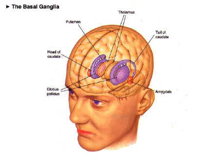
Anatomy/functionality
Today we are going to cover the Basal ganglia.
The functions of the Basal ganglia are as follows:
*Controls Cognition
*Movement Coordination
*Voluntary Movement
More to come
Today we are going to cover the Basal ganglia.
The basal ganglia is located deep within the cerebral hemispheres in the telencephalon region of the brain. It consists of the corpus stratium,subthalamic nucleus and the substantia nigra.
Source: biology.about.com
The functions of the Basal ganglia are as follows:
*Controls Cognition
*Movement Coordination
*Voluntary Movement
More to come
Hey guys, I've been practising Reiki daily for quite some time now. It would be interesting to be hooked up to a machine when I commence healing and
see what type of brain wave, etc eminate from me. Do any of you have an idea of how this could be accomplished?
I would upload my course notes for each type of Reiki, but the files are too big. Does anyone have a site I could upload them to so I will be able to share them?
Thanks,
bs
I would upload my course notes for each type of Reiki, but the files are too big. Does anyone have a site I could upload them to so I will be able to share them?
Thanks,
bs
Ok, long time no see...I've been going through a few bouts with my girlfriend and didn't want to depress any of you guys .
Reiki...where to start...
When I first started using it, I wasn't really sure what I was doing. As I got more into it, I started visualizing energy surrounding me and sometimes I would enter a meditative state while healing myself. I have healed others, but the problem is they have to be able to "accept" the energy or it will not heal them in the proper manner. However, once a person/myself has accepted it into their life, the energy knows exactly which channels to go through and heals automatically. I do not have to tell it to heal the head injury, etc. It will merely find the energy field limited in certain areas and fill in the blanks. I have been given this analogy to use before: Compare the body to a few potholes on a sidewalk. If you drop a bucket of water on it, does the water not go to the lowest point? This is the same with energy. It will heal whatever is needing the energy, not overextend other parts of the body.
Picture a shimmering bright light around everyone that can fluctuate with moods, actions, etc. What we do, say, feel, and interpret will change the aura field around us. Occassionally, this aura field becomes emotionally damaged due to something happening to us. The aura will heal itself slowly, but that part may never gain the same amount of energy it had before. Sometimes people have called this the pain body, but that has to deal with the egoic storage of past experiences. If you are able to release these experiences, your auric field will be very healthy and not hinder on your life. If you have a large ego, (for instance) your past will follow you wherever you go. It is possible to heal this through Reiki, but I would suggest Reiki only to be a helping hand. Any severe psychological work should be done in tandem witht his sort of healing. You don't want to end up schizophrenic do ya? (I don't think that would be possible, but you get my drift I hope).
Any further questions? I'd be glad to help.
New horizons in "F"MRI
Pioneering steps towards understanding the "true" Final Frontier..
Semper
Spin-Echo Magnetic Resonance Imaging
In spin-echo MRI, gradients and Fourier analysis are used to perform three-dimensional imaging. Other techniques of MRI, such as gradient-echo, are slight variations of spin-echo imaging, so I will only describe spin-echo imaging in detail. The component of the imaging system which allows the spatial localization of the protons is a set of magnetic field gradients, set up by magnetic coils which are turned on and off at appropriate times (Horowitz, 1995).
When hydrogen nuclei relax, the frequency that they transmit is positively correlated with the strength of the magnetic field surrounding them. A magnetic field gradient along the z-axis, called the "slice select gradient," is set up when the RF pulse is applied, and is shut off when the RF pulse is turned off. This gradient causes the hydrogen nuclei at the high end of the gradient (where the magnetic field is strong) to precess at a high frequency (e.g., 65 MHz), and those at the low end (weak field) to precess at a lower frequency (e.g., 63 MHz). When the RF pulse, of a single frequency, is applied, only those nuclei which precess at that frequency will be tilted, to later relax and emit a radio transmission (i.e., the nuclei "resonate" to that frequency). For example, if the magnetic gradient caused hydrogen nuclei to precess at rates from 63 MHz at the low end of the gradient to 65 MHz at the high end, and the gradient were set up such that the high end was located at the patient's head and the bottom part at the patient's feet, then a 63 MHz RF pulse would excite the hydrogen nuclei in a slice near the feet, and a 65 MHz pulse would excite them in a slice near the head. Thus a single "slice" along the z-axis is selected; only the protons in this slice are excited to a higher energy level, to later relax to a lower energy level and emit a radio transmission (Horowitz, 1995).
The second dimension of the image is extracted with the help of a phase encoding gradient. Immediately after the RF pulse ceases, all of the nuclei in the activated slice are "in phase," that is, their magnetic vectors all point in the same direction. Left to their own devices, these vectors would relax. In MRI, however, the phase encoding gradient (in the y-dimension) is briefly applied, in order to cause the magnetic vectors of nuclei along different portions of the gradient to point in different directions (Horowitz, 1995).
After the RF pulse, slice select gradient, and phase encoding gradient have been turned off, the MRI instrument sets up a third magnetic field gradient, along the x axis, called the "frequency gradient" or "read-out gradient." This gradient causes the relaxing protons to be differentially re-excited, so that the nuclei near the low end of the gradient begin to precess at a faster rate, and those at the high end pick up even more speed. When these nuclei relax again, the fastest ones (those which were at the high end of the gradient) will emit the highest frequency of radio waves. The frequency gradient is applied "only when the signal is measured" (Horowitz, 1995).
The second and third dimensions of the image are extracted by means of Fourier analysis. The entire procedure must be repeated multiple times in order to form an image with a good signal-to-noise ratio.
Finally, in spin-echo imaging, there is the problem that the inhomogeneity of the main magnetic field induces variations in the rate of precession of nuclei. To fix this problem, a 180-degree RF pulse is inserted into the cycle, at a time point halfway between the 90-degree pulse and the measurement of the radio transmission signal given off by the relaxing nuclei (Horowitz, 1995).
www.neuroguide.com...
Pioneering steps towards understanding the "true" Final Frontier..
Semper
new topics
-
Weinstein's conviction overturned
Mainstream News: 52 minutes ago -
Supreme Court Oral Arguments 4.25.2024 - Are PRESIDENTS IMMUNE From Later Being Prosecuted.
Above Politics: 2 hours ago -
Krystalnacht on today's most elite Universities?
Social Issues and Civil Unrest: 2 hours ago -
Chris Christie Wishes Death Upon Trump and Ramaswamy
Politicians & People: 2 hours ago -
University of Texas Instantly Shuts Down Anti Israel Protests
Education and Media: 5 hours ago -
Any one suspicious of fever promotions events, major investor Goldman Sachs card only.
The Gray Area: 7 hours ago -
God's Righteousness is Greater than Our Wrath
Religion, Faith, And Theology: 11 hours ago
top topics
-
VP's Secret Service agent brawls with other agents at Andrews
Mainstream News: 16 hours ago, 11 flags -
Krystalnacht on today's most elite Universities?
Social Issues and Civil Unrest: 2 hours ago, 7 flags -
Nearly 70% Of Americans Want Talks To End War In Ukraine
Political Issues: 17 hours ago, 6 flags -
Sunak spinning the sickness figures
Other Current Events: 16 hours ago, 5 flags -
Supreme Court Oral Arguments 4.25.2024 - Are PRESIDENTS IMMUNE From Later Being Prosecuted.
Above Politics: 2 hours ago, 5 flags -
Weinstein's conviction overturned
Mainstream News: 52 minutes ago, 4 flags -
Electrical tricks for saving money
Education and Media: 14 hours ago, 4 flags -
University of Texas Instantly Shuts Down Anti Israel Protests
Education and Media: 5 hours ago, 2 flags -
Any one suspicious of fever promotions events, major investor Goldman Sachs card only.
The Gray Area: 7 hours ago, 2 flags -
Chris Christie Wishes Death Upon Trump and Ramaswamy
Politicians & People: 2 hours ago, 1 flags
active topics
-
Chris Christie Wishes Death Upon Trump and Ramaswamy
Politicians & People • 9 • : Hecate666 -
Nearly 70% Of Americans Want Talks To End War In Ukraine
Political Issues • 80 • : FlyersFan -
Supreme Court Oral Arguments 4.25.2024 - Are PRESIDENTS IMMUNE From Later Being Prosecuted.
Above Politics • 29 • : xuenchen -
University of Texas Instantly Shuts Down Anti Israel Protests
Education and Media • 103 • : DBCowboy -
VP's Secret Service agent brawls with other agents at Andrews
Mainstream News • 45 • : network dude -
Weinstein's conviction overturned
Mainstream News • 10 • : xuenchen -
Remember These Attacks When President Trump 2.0 Retribution-Justice Commences.
2024 Elections • 57 • : TzarChasm -
-@TH3WH17ERABB17- -Q- ---TIME TO SHOW THE WORLD--- -Part- --44--
Dissecting Disinformation • 670 • : cherokeetroy -
British TV Presenter Refuses To Use Guest's Preferred Pronouns
Education and Media • 159 • : 5thHead -
HORRIBLE !! Russian Soldier Drinking Own Urine To Survive In Battle
World War Three • 40 • : Myhandle