It looks like you're using an Ad Blocker.
Please white-list or disable AboveTopSecret.com in your ad-blocking tool.
Thank you.
Some features of ATS will be disabled while you continue to use an ad-blocker.

The Illusion of Color: Perceptions of Light
page: 13
share:
Hi all, I've recently prepared a series of slides, some of which I've made into a mini-presentation that I thought might be interesting to the folks
here on ATS. The subject presented here is the nature of light, and how we perceive it; the broader topic is information loss in human systems of
perception.
I thought this might be interesting because people really don't seem to understand the difference between color and light, and how our human-specific systems of perception work. This is an important thing to understand, especially when understanding spectroscopy and 'false color' images, for example those which NASA releases.
Anyway, I've resized some slides and written some language around them; hope you enjoy and perhaps find it informative.
A Brief Overview Of Color
Our eyes react to light, which we perceive as having various 'colors'. It's important to realize that 'color' is not a property of the light itself, it's a property of our perception of the light.
Light, being electromagnetic radiation, can be at different wavelengths, and with different energy levels at each wavelength. The light we see is almost always a blend of different wavelengths of photons, in various proportions, ranging from about 400 nanometers to 700 nanometers (the 'visible spectrum').
The Cells Of The Retina
The retina is the portion of the eye that reacts to light. Cells on the retina are stimulated by photons, build up an electrical potential, and transmit 'activation' signals to other nerve cells in the eye, eventually transmitting the information to the visual cortex.
There are two major categories of light-sensitive cells in the retina: rods and cones (named for their rough shapes). Rod cells are more sensitive to light intensity, along a wider range of wavelengths, than cone cells. They're the primary cell responsible for night-vision and monochrome perception. Cone cells are responsible for color vision.
The different types of cells are distributed in varying densities along the surface of the retina. The areas of the retina that generate peripheral vision contain mostly rod cells, while the center of the retina (the fovea) is mostly cone cells. This explains why, when it's very dark, you can sometimes see dimly-lit objects with better contrast by looking to one side or the other, instead of staring straight at them.
The cone cells are of three types: known as S-cones, M-cones, and L-cones. Each type responds differently to different wavelengths of light. They're sometimes called blue, green, and red cones, but that labeling isn't really accurate -- remember, wavelengths aren't quite the same thing as colors.
The M-Cone And Monovariant Color Perception
Let's look first at M-cones (the 'green' receptors of the eye). When photons are absorbed by a cone, they cause that cone to build up an electrical potential -- the 'signal'. Due to the type of photosensitive molecule in the cone, it gets more or less potential built-up from different wavelengths of photons. Here's a graph of the M-cone activation response:
Let's look at how two different samples of pure-wavelength light affect the M-cone. The first sample, 'A', is at 485nm, the second, 'B', is at 608nm:
It's important to notice that even though these samples each have the same number of photons per second impacting the retina, they result in different activation energy levels from the cone. Clearly, a field of L-cones can distinguish between 'A' and 'B'. But what if we 'turn up the brightness' of 'B', doubling the number of photons per second?
So a color perception system with a single type of 'sensor' can be quite easily fooled!
The L-Cone And Divariant Color Perception
The second type of cone in the eye is called the L-cone (or 'red' receptor). It has a different activation graph than the M-cone, as it uses a slight variant of the same photosensitive chemical:
This different activation curve allows the L-cone to quite neatly detect differences that the M-cone misses, and vice-verse. For example, the previous examples of 'A' versus 'B' and 'A' versus '2 * B', in terms of the L-Cone response:
But can a divariant color perception system still be fooled? Yep! Let's add another pure-wavelength of light, 'C', at 580nm:
While that's obviously distinguishable from either 'A' or 'B', lets look at how it is perceived versus the blend of 'A + B':
The S-Cone And Trivariant Color Perception
The normal human eye has three types of cone cells. The third type is known as the S-cone, it has a much lower range of wavelength activation than the other two types of cones; it is more sensitive in the 'blue range':
Here's what the wavelengths 'A', 'B', and 'C' look like, with the S-cone activatation curve added:
This obviously clears up the situation that was indistinguishable with only a two-cone system, as only wavelength 'A' causes any activation energy in the S-cone:
Trivariant Color Aliasing
But can a trivariant system, with all three different types of cones, be fooled? Of course! For example, considering the following, with new locations for 'B' (at 412nm) and 'C' (at 520nm):
The trick is to remember that different wavelengths of light can be blended in different proportions. Here's a comparison of 'A' with 'B + 2/5 * C':
These two very different compositions of light are perceived by humans as virtually identical colors.
Enough for now; hope this was interesting and/or informative!
sweet...I was looking at this right before I saw and clicked this thread
I sometimes ask a question from people, just for fun and for a hope that somebody understands out of human perception.
As child, you are taught what color is which. Red, for example. Then you go and see that wavelength, and you say it is red. You can also distinguish blue.
But what if humans are simply taugth to realize that it does not matter what they perceive, even if they see red as blue and blue as red, since they can perfectly adapt into that situation?
Does that even matter? It is truth, tough.
reply to post by rawsom
Actually, it's not true. Now, if everyone called red 'blue' and blue 'red', then yes, it would make no difference. After all, what's in a name?
But if you consider the wavelengths of light involved in those colors, and the types of sensors the eye uses, red and blue are not interchangeable.
Light around 450nm (blue) is much more susceptible to chromatic aberration effects, for this reason the very center of the fovea contains almost no S-cones, in order that the eye may better detect fine detail. There's also the 'blue-field entoptic' effect, which can often be seen when staring at a bright blue sky. That doesn't happen with red.
The output of the cones in the eye is processed by a series of cells known as 'color opponent cells', which balance contrast between various combinations of cone-type inputs. The most common type balances G+/R-, or G-/R+. Blue is treated quite differently, usually contrasting with the combination or absence of green and red, eg B+/(G+R)- or B-/(G+R)+.
reply to post by iiinvision
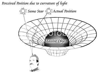
iiinvision, a very interesting image to ponder. Another way of looking at it: light takes the path of least time between any two points, regardless of whether that is a straight line or not. If mass density is considered a non-homogenous addition to the 3 spatial dimensions, that path can be considered as traveling along the surface of a higher-dimensional solid, similar to how the shortest path between two points along the surface of the Earth is an arc, not a straight line.
iiinvision, a very interesting image to ponder. Another way of looking at it: light takes the path of least time between any two points, regardless of whether that is a straight line or not. If mass density is considered a non-homogenous addition to the 3 spatial dimensions, that path can be considered as traveling along the surface of a higher-dimensional solid, similar to how the shortest path between two points along the surface of the Earth is an arc, not a straight line.
Forgot to cite my data source... even though the graphs may look hand-drawn, they're not. I plotted them using an OpenOffice spreadsheet.
The source data is taken from the CVRL dataset:
10-deg fundamentals based on the Stiles and Burch 10-deg CMFs
The data is precise to 1nm increments, with a linear representation of energy response.
The source data is taken from the CVRL dataset:
10-deg fundamentals based on the Stiles and Burch 10-deg CMFs
The data is precise to 1nm increments, with a linear representation of energy response.
new topics
-
whistleblower Captain Bill Uhouse on the Kingman UFO recovery
Aliens and UFOs: 6 minutes ago -
1980s Arcade
General Chit Chat: 2 hours ago -
Deadpool and Wolverine
Movies: 3 hours ago -
Teenager makes chess history becoming the youngest challenger for the world championship crown
Other Current Events: 4 hours ago -
CIA botched its handling of sexual assault allegations, House intel report says
Breaking Alternative News: 5 hours ago -
Lawsuit Seeks to ‘Ban the Jab’ in Florida
Diseases and Pandemics: 7 hours ago -
Starburst galaxy M82 - Webb Vs Hubble
Space Exploration: 9 hours ago -
15 Unhealthiest Sodas On The Market
Health & Wellness: 9 hours ago -
The Superstition of Full Moons Filling Hospitals Turns Out To Be True!
Medical Issues & Conspiracies: 11 hours ago
top topics
-
Lawsuit Seeks to ‘Ban the Jab’ in Florida
Diseases and Pandemics: 7 hours ago, 18 flags -
Starburst galaxy M82 - Webb Vs Hubble
Space Exploration: 9 hours ago, 11 flags -
The Superstition of Full Moons Filling Hospitals Turns Out To Be True!
Medical Issues & Conspiracies: 11 hours ago, 8 flags -
CIA botched its handling of sexual assault allegations, House intel report says
Breaking Alternative News: 5 hours ago, 8 flags -
IDF Intel Chief Resigns Over Hamas attack
Middle East Issues: 14 hours ago, 6 flags -
15 Unhealthiest Sodas On The Market
Health & Wellness: 9 hours ago, 5 flags -
It takes One to Be; Two to Tango; Three to Create.
Philosophy and Metaphysics: 15 hours ago, 4 flags -
Deadpool and Wolverine
Movies: 3 hours ago, 3 flags -
1980s Arcade
General Chit Chat: 2 hours ago, 3 flags -
Teenager makes chess history becoming the youngest challenger for the world championship crown
Other Current Events: 4 hours ago, 2 flags
3