It looks like you're using an Ad Blocker.
Please white-list or disable AboveTopSecret.com in your ad-blocking tool.
Thank you.
Some features of ATS will be disabled while you continue to use an ad-blocker.

Your Doctor Can Now Examine an Exact 3D Replica of Your Heart in Virtual Reality
page: 14
share:
Stumbled upon this today.
I think this is great step forwards in medical science. This new innovation is exactly as the title suggests. 3D mapping your Heart for viewing. This will lead to a ton of fantastic applications and hopefully many saved lives.
They are already applying this to other organs as well, as suspected. Hopefully, we'll see an increase in identification and successful surgery rates with this easy-to-spot system too!
We see a huge opportunity for this sort of imaging technology to shape the way doctors work.
Source
Here's for a better future!
I think this is great step forwards in medical science. This new innovation is exactly as the title suggests. 3D mapping your Heart for viewing. This will lead to a ton of fantastic applications and hopefully many saved lives.
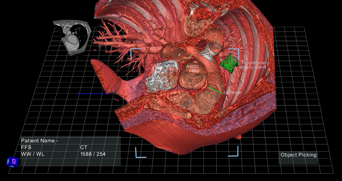
Today, radiologists look through hundreds and hundreds of flat images, and then draw a diagram (yes, by hand) to show the surgeon how to approach a given procedure. Then the surgeon operates on the patient with no advance knowledge of his or her actual volumetric anatomy.
One surgeon I spoke with summed it up perfectly: “I’ve never opened up a patient and seen a 2D view!” Another surgeon specializing in image-guided surgery told us that “half the time I am guessing” when navigating 3D anatomy using 2D images.
Sounds a little scary, no? Never fear. It won’t be like this much longer.
There’s a pretty powerful solution just now arriving—advanced image rendering through interactive virtual reality. EchoPixel (my company) uses virtual reality to help doctors visualize each patient’s unique anatomy and internal structure in a floating 3D image. The software uses DICOM data, which is already embedded in every MRI scan, CT scan, or ultrasound image.
They are already applying this to other organs as well, as suspected. Hopefully, we'll see an increase in identification and successful surgery rates with this easy-to-spot system too!
We see a huge opportunity for this sort of imaging technology to shape the way doctors work.
We see a huge opportunity for this sort of imaging technology to shape the way doctors work.
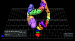
For example, the virtual colonoscopy procedure is becoming a popular alternative to the dreaded optical colonoscopy (recommended as a regular procedure for every person over age 50). Instead of requiring total sedation and a full day of recovery, the virtual procedure allows the doctor to examine a CT scan of the colon to identify any potentially cancerous lesions.
Source
Here's for a better future!
a reply to: Ghost147
Does this replace the torturous T.E.E. ?
Having had a TEE , transesophigeal echocardiogram, done not long ago, this news is, as the younger set say, *a total bummer* for me. If you've ever had the equivalent of a broom handle wrapped with electrical tape and a small marble mounted on the end SHOVED down your throat, while awake, then you *catch my drift *.
(That numbing goop they insist numbs your throat...doesn't.)
To those of you who can benefit from this technology, I am genuinely happy for you!
new topics
-
Has Tesla manipulated data logs to cover up auto pilot crash?
Automotive Discussion: 33 minutes ago -
whistleblower Captain Bill Uhouse on the Kingman UFO recovery
Aliens and UFOs: 5 hours ago -
1980s Arcade
General Chit Chat: 7 hours ago -
Deadpool and Wolverine
Movies: 8 hours ago -
Teenager makes chess history becoming the youngest challenger for the world championship crown
Other Current Events: 9 hours ago -
CIA botched its handling of sexual assault allegations, House intel report says
Breaking Alternative News: 10 hours ago
4